Microscopy
只今準備中
顕微鏡で見えたものを、少しずつ並べていきます
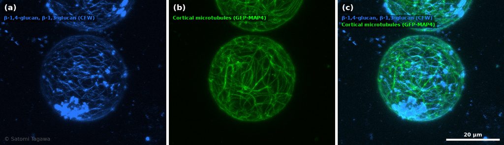
顕微鏡による可視化を、これまで研究の軸に据えてきました。
論文として発表してきた画像の中から、代表的なものをこれから少しずつご紹介していく予定です。
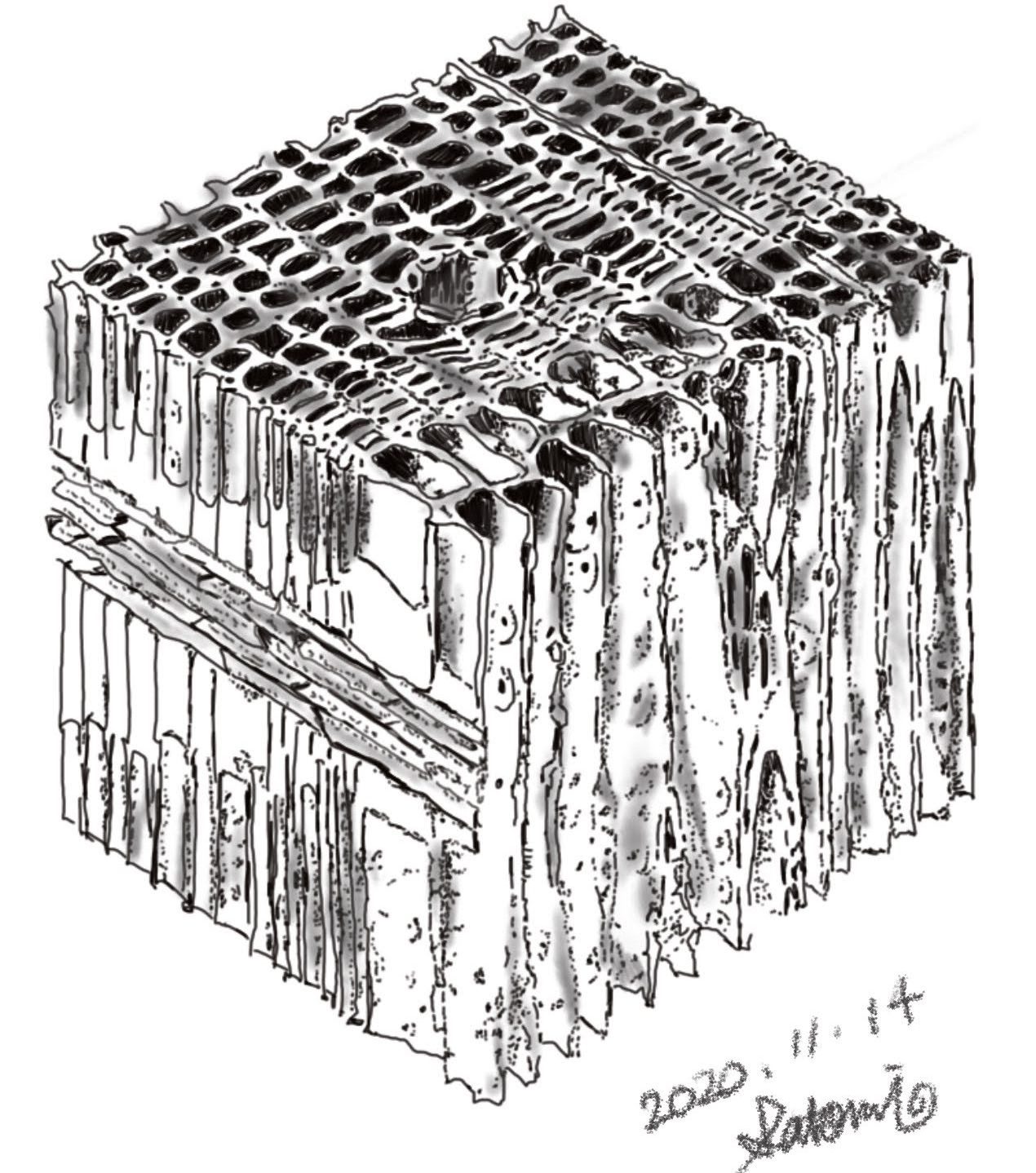
素材そのものを手に取って眺めても、内部で何が起きているのかはほとんど分かりません。前処理を工夫し、染め方や光の当て方を変え、倍率を上げていき、そうしてはじめて、構造が姿を現します。これから並べていく一枚一枚は、その瞬間を切り取ったものですっ!
論文の中では図の一つに過ぎませんが、どの画像にも撮影までの試行錯誤があり、見えたときの驚きがあります。そうした背景も少し添えながら、順に掲載していきます。
Sharing What the Microscope Has Shown Me
Visualization through microscopy has been at the core of my research. Over the coming posts, I plan to share some of the representative images from the papers I have published.
Looking at a material with the naked eye tells you almost nothing about what is happening inside it. It is only after refining the sample preparation, changing the staining and the way light falls on it, and pushing the magnification higher that the structure finally reveals itself. Each image I will be posting here captures one of those moments.
Within a paper, an image is just one figure among many. But behind every one of them lies a good deal of trial and error, and the small shock of finally seeing something. I will add a little of that background as I go, and post them one by one.